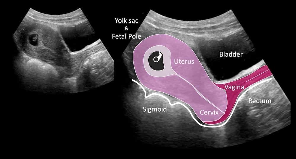

In the third trimester, ultrasound is used to assess fetal growth and well-being based upon various clinical indications to be reviewed with individual care providers. In the second trimester, ultrasound imaging is used to evaluate the fetal anatomy and possibly to evaluate the cervical length as a marker of risk for pre-term birth often based upon certain risk factors. Finally, screening for chromosomal/genetic problems can be provided to patients if desired through various nuchal translucency screening programs. Most importantly, it confirms the gestational age of the pregnancy to allow for proper timing of care throughout the remainder of pregnancy.

In the first trimester, ultrasound is used to identify the location, viability, and number of fetuses present. There are no known or proven harmful effects of medical diagnostic ultrasound to human fetuses when using standard clinical settings on the machines. The handheld device both emits and detects these sound waves, measuring the time it took each wave to be reflected back to the handpiece.īecause certain tissues, like bone, fluid, and soft tissue, reflect sound differently than others, certain boundaries can be identified in the timing of reflected ultrasound pulses. This is what creates the image you and the physician see in the ultrasound monitor, or sonogram. Ultrasound imaging devices work by emitting high-frequency sound waves (far above the threshold for human hearing) that are reflected by the tissues of the human body. Ultrasound is generally considered safe in pregnancy for medical diagnostic purposes with focused evaluations. However, this does not mean any and every ultrasound procedure is recommended, such as so-called “keepsake” ultrasound images and video. The safety of unnecessary prolonged ultrasound exposure for entertainment purposes has not been well studied in in the medical literature and is not currently advocated by national organizations (FDA, AIUM, ACOG, SMFM). Ultrasound is the most commonly used medical imaging technology in pregnancy to view the unborn fetus and determine the health of the pregnancy. Throughout each trimester, ultrasound imaging, also called sonography, is used to ensure both the mother and fetus are doing well. Current ultrasound technology allows for traditional two-dimensional (2D) imaging as well as the more recent 3D and 4D (or 3D video) evaluations.


 0 kommentar(er)
